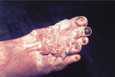
A 55-year-old man presented with vegetating lesions on the right foot that had been slowly enlarging during the past several years ;The photo:
On physical examination, several nodular and verrucous lesions were seen in the distal region of the foot. The patient lived in a rural area and had walked barefoot for most of his life. Analysis of a skin-biopsy specimen revealed clusters of small, round, thick-walled, brown sclerotic bodies in the stratum corneum (muriform cells), which are diagnostic for chromoblastomycosis.
Chromoblastomycosis is a chronic, soft-tissue fungal infection commonly caused by Fonsecaea pedrosoi, Phialophora verrucosa, Cladosporium carrionii, or F. compacta. The infection occurs in tropical or subtropical climates and often in rural areas. The fungi are usually introduced to the skin through cutaneous injury from thorns, splinters, or other plant debris. The patient was treated with multiple surgical excisions and itraconazole for 24 months with a complete resolution of symptoms.

Custom Search
Popular Posts
-
W ind --- pneumonia, atelectasis at 1st 24- 48 hours W ater --- urinary tract infection at Anytime after post op day 3 W ound --- wound ...
-
Raccoon Eyes. Ecchymosis in the periorbital area, resulting from bleeding from a fracture site in the anterior portion of the skull base. Th...
-
Lymph Nodes : The major lymph node groups are located along the anterior and posterior aspects of the neck and on the underside of the jaw. ...
-
Fournier's gangrene is a rare condition and delayed treatment results in fatal outcome. We managed a case of Fournier's gangrene by...
-
Viewed posteriorly the right kidney has its upper edge opposite the 11th dorsal spine and the lower edge of the 11th rib. Its lower edge is ...
-
If you are palpating a swelling like an abdominal swelling infront of the aorta, You have to decide whether the mass you feel is pulsatile/e...
-
The sphenoid bone carries its share of creating part of the base of the cranium. While it can be seen laterally and inferiorly, the shape of...
-
Look at how your jaw ends up when saying first syllable of 'Lateral' or 'Medial' : - "La" : your jaw is now open ...


